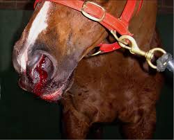
Managing the horse with internal haemorrhage is challenging but can be rewarding.

The medical records of 19 horses with acute hemoperitoneum were reviewed. The causes for the hemoperitoneum were idiopathic (8 horses), splenic hematoma with capsular tear (7), bleeding from the reproductive tract (3), multicentric hemangiosarcoma (1), and systemic amyloidosis (1). The affected horses were between 4 and 32 years of age (median 11.5 years). The most consistent findings on initial examination were depression, tachycardia, tachypnea, pale mucous membranes, prolonged capillary refill time, colic, and abdominal discomfort. Less common clinical signs included abdominal distention, profuse sweating, ataxia, and broad ligament mass palpated on rectal examination. Clinicopathologic abnormalities commonly detected were anemia, neutrophilia, lymphopenia, thrombocytopenia, hypoproteinemia, hypocalcemia, azotemia, increased creatinine kinase, and sorbitol dehydrogenase activity. Hemoperitoneum was diagnosed on the basis of abdominocentesis, transabdominal ultrasonography, and postmortem examination. Sixteen horses were treated, and 3 horses were euthanized at owners' request because of severe clinical signs. The treatment consisted of the administration of intravenous fluids, plasma or blood transfusion, nonsteroidal drugs, antimicrobial drugs, and antifibrinolytic and procoagulant agents. Rapid clinical deterioration was observed in 2 horses, necessitating euthanasia. The remaining 14 horses survived the abdominal bleeding (survival rate 74%) and were discharged 3–15 days (median 7.0 days) after presentation. Postmortem examination of the 6 nonsurvivors showed massive abdominal hemorrhage from splenic hematoma with capsular tear (2 horses), multicentric hemangiosarcoma with liver rupture (1), systemic amyloidosis with splenic hematoma and capsular tear (1), and bilateral ruptured ovarian hematomas (1). In one horse, no origin of the bleeding could be determined during postmortem examination.
Click here to be directed to the study in the Journal of Veterinary Internal Medicine.

